Categories more
- Adventures (62)
- Arts / Collectables (14)
- Automotive (77)
- Aviation (65)
- Bath, Body, & Health (123)
- Children (6)
- Cigars / Spirits (71)
- Cuisine (66)
- Design/Architecture (22)
- Electronics (63)
- Entertainment (36)
- Event Planning (115)
- Fashion (45)
- Finance (10)
- Gifts / Misc (6)
- Home Decor (118)
- Jewelry (41)
- Pets (3)
- Philanthropy (1)
- Real Estate (16)
- Services (44)
- Sports / Golf (14)
- Vacation / Travel (85)
- Watches / Pens (33)
- Wines / Vines (34)
- Yachting / Boating (67)


Innovations in Advanced Urological Care and Treatments

Published
08/19/2026Diagnostic Evolution: The Role of TRUS in Advanced Urological Care
The landscape of urological diagnostics has undergone significant evolution, with Transrectal Ultrasound (TRUS) playing a pivotal role. This technology leverages sound waves to generate real-time images, offering clinicians a dynamic view of the prostate and surrounding pelvic structures. Often referred to as an endorectal ultrasound or prostate sonogram, TRUS has become a cornerstone in evaluating prostate health, guiding interventions, and enhancing diagnostic precision.
Mechanism and Application of Transrectal Ultrasound
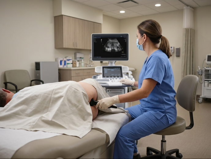
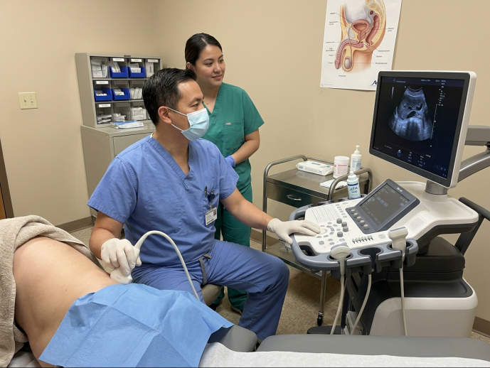
At its core, TRUS operates on the principle of sonography. A small, finger-sized probe, known as a transducer, is gently inserted into the rectum. This transducer emits high-frequency sound waves that travel through tissues, bouncing back as echoes. A computer then processes these echoes, converting them into a detailed image displayed on a monitor. This real-time visualization allows urologists to assess organ structure, size, and any anomalies.
To optimize image quality, modern TRUS systems offer advanced settings. Clinicians can adjust the dynamic range, for instance, to enhance image contrast, making subtle tissue variations more apparent. While harmonic imaging provides higher spatial resolution, fundamental imaging often offers superior acoustic density and contrast, which can be crucial for distinguishing between normal and abnormal tissue based on their echogenicity patterns. Peripheral zone cancers, for example, frequently appear as hypoechoic (darker) lesions, while transition zone cancers might present as hyperechoic (brighter) masses. This ability to fine-tune imaging parameters transforms TRUS from a mere navigation tool into a powerful diagnostic modality.
TRUS vs. Multiparametric MRI in Modern Diagnostics
When considering prostate imaging, TRUS and multiparametric MRI (mpMRI) are frequently discussed. Both offer unique advantages, and their roles are often complementary rather than mutually exclusive.
Multiparametric MRI (mpMRI) is renowned for its superior soft-tissue resolution and ability to provide detailed anatomical and functional information. It excels at distinguishing healthy prostate tissue from cancerous lesions and is often used for risk stratification, particularly with the PI-RADS (Prostate Imaging-Reporting and Data System) scoring system. mpMRI can detect lesions that might be "invisible" on standard TRUS, especially in the anterior prostate.
However, TRUS holds distinct advantages in terms of accessibility, cost efficiency, and real-time visualization. TRUS equipment is widely available in most urology clinics, making it a more immediate and less expensive option than mpMRI. Its real-time capability is invaluable for guiding procedures directly. While mpMRI provides a detailed static map, TRUS offers dynamic guidance during biopsies or other interventions. Furthermore, TRUS is crucial for accurate PSA density calculation, a metric derived by dividing the blood PSA level by the prostate volume (measured via TRUS), which helps differentiate benign prostatic hyperplasia (BPH) from prostate cancer.
The choice between TRUS and mpMRI often depends on the clinical context. For initial prostate evaluation, TRUS is frequently the first line due to its convenience and lower cost. For suspicious findings or when greater detail is required, mpMRI provides an excellent complementary view. In many modern practices, mpMRI is performed before a biopsy, and TRUS is then used to guide the biopsy needle to the specific areas identified by the MRI, sometimes using software-based fusion.
Feature Transrectal Ultrasound (TRUS) Multiparametric MRI (mpMRI) Availability Widely available in clinics Less widely available, often requires specialized centers Cost Lower Higher Real-time Imaging Yes, excellent for guidance No, provides static images Resolution Good, especially with optimized settings Excellent, superior soft-tissue detail Radiation Exposure None (uses sound waves) None (uses magnetic fields and radio waves) Primary Use Biopsy guidance, volume measurement, initial assessment Cancer detection, staging, risk stratification (PI-RADS) Accessibility Quick, in-office procedure Longer scan time, requires specialized equipment Diagnostic Output Echogenicity patterns, prostate size, PSA density Detailed anatomical, functional, and metabolic information
Clinical Indications and Conditions Managed Through TRUS
TRUS is a versatile tool with a broad spectrum of clinical indications, ranging from routine prostate evaluations to guiding complex therapeutic interventions. Its ability to provide clear visualization of pelvic organs makes it indispensable in modern urology.
Diagnosing Prostate Cancer, BPH, and Pelvic Conditions
One of the primary applications of TRUS is the detailed assessment of the prostate gland. It allows clinicians to accurately measure prostate size, which is critical for diagnosing and managing benign prostatic hyperplasia (BPH), a common condition affecting men as they age. By calculating prostate volume, TRUS helps determine appropriate treatment strategies, from medication to surgical interventions.
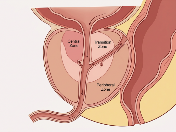
Beyond size, TRUS provides insights into the prostate's internal architecture, allowing for the differentiation of its zonal anatomy—specifically the peripheral zone and transition zone. Most prostate cancers originate in the peripheral zone, which TRUS can effectively image. While not all prostate cancers are clearly visible on TRUS, suspicious hypoechoic areas can be identified and targeted for biopsy. TRUS is also valuable in diagnosing prostatitis (inflammation of the prostate) and identifying other pelvic conditions such as cysts in the seminal vesicles or prostatic and pelvic abscesses. Its utility extends to evaluating male infertility by examining the seminal vesicles and ejaculatory ducts for obstructions or abnormalities.
Here is a list of common pelvic conditions that TRUS can help diagnose:
- Benign Prostatic Hyperplasia (BPH)
- Prostate Carcinoma (cancer)
- Prostatitis (prostate inflammation)
- Prostatic Cysts
- Seminal Vesicle Cysts or Inflammation
- Ejaculatory Duct Obstruction
- Pelvic Abscesses (e.g., prostatic, perirectal)
- Male Infertility (evaluating reproductive structures)
Therapeutic Interventions and Image-Guided Procedures
The real-time imaging capability of TRUS makes it an excellent tool for guiding various therapeutic interventions. The most common guided procedure is the needle core biopsy, where TRUS precisely directs the biopsy needle to suspicious areas within the prostate, ensuring accurate tissue sampling for pathological analysis. This guidance is crucial for confirming a prostate cancer diagnosis and determining its aggressiveness.
Beyond diagnostics, TRUS facilitates several advanced treatments for prostate cancer:
- Brachytherapy Placement: TRUS guides the precise implantation of radioactive seeds into the prostate gland, delivering targeted radiation therapy.
- Cryotherapy Guidance: It helps accurately position probes that freeze and destroy cancerous tissue within the prostate.
- High-Intensity Focused Ultrasound (HIFU) Treatment: TRUS provides real-time monitoring during HIFU, ensuring the focused ultrasound energy ablates only the targeted cancerous areas.
- Abscess Aspiration: For patients with prostatic or pelvic abscesses, TRUS can guide a needle to drain the fluid, alleviating symptoms and aiding recovery.
These image-guided procedures underscore TRUS's role in delivering highly targeted and effective treatments, contributing significantly to the personalized urology services that are the hallmark of modern medical care. For individuals seeking comprehensive and tailored urological solutions, understanding these advanced techniques is key to informed decision-making.
Comparative Analysis: TRUS-Guided Biopsy vs. Transperineal Approaches
When a prostate biopsy is necessary, two primary approaches are commonly employed: transrectal ultrasound (TRUS)-guided biopsy and transperineal biopsy. Both aim to obtain tissue samples for cancer diagnosis, but they differ significantly in their procedural nuances, associated risks, and effectiveness.
Procedural Differences, Accuracy, and Infection Control
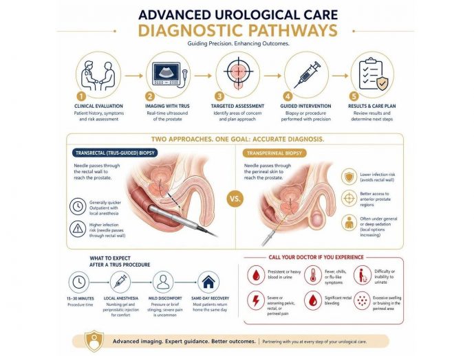
The TRUS-guided biopsy involves passing a needle through the rectal wall into the prostate gland to collect tissue samples. This transrectal sampling method is often quick, convenient, and performed with local anesthesia. Historically, it has been the standard of care for prostate biopsies. However, a significant concern with the TRUS approach is the risk of infection. The needle passes through the rectal flora, which naturally contains bacteria, leading to a potential for introducing these bacteria into the prostate and bloodstream. Post-biopsy infection rates have risen, partly due to increasing fluoroquinolone resistance among common rectal bacteria. This can result in complications ranging from urinary tract infections to severe sepsis, which can be life-threatening.
In contrast, the transperineal biopsy involves inserting the biopsy needle through the skin of the perineum (the area between the scrotum and anus), directly into the prostate. This approach avoids passing through the rectal wall, significantly reducing the risk of infection by bypassing the rectal flora. While typically performed under general anesthesia or deep sedation, advancements are making local anesthesia more feasible.
Regarding diagnostic accuracy, both methods can be effective, especially when guided by pre-biopsy mpMRI. However, transperineal biopsy often offers better access to the anterior prostate, where some cancers can be missed by the transrectal approach. For pain management, periprostatic nerve blockade, involving the injection of local anesthetic around the prostate, is routinely used for TRUS biopsies to minimize discomfort.
A key statistic highlights the difference in complications: TRUS biopsy is associated with post-biopsy infection rates rising from approximately 1% historically to between 2–4% in recent years. This contrasts with transperineal biopsy, which generally has lower infection rates. Conversely, TRUS biopsy typically carries a lower risk of acute urinary retention (0.4–6%) compared to transperineal biopsy (1.7–11.1%), primarily because the transperineal approach often involves more extensive needle passes through the prostate tissue, leading to more swelling.
Integrating Advanced Urological Care with Targeted Prophylaxis
To mitigate the infection risks associated with TRUS-guided biopsies, advanced urological care emphasizes rigorous infection control strategies. A crucial step is the implementation of targeted antibiotic prophylaxis. This involves performing pre-biopsy rectal swabs to culture the patient's rectal flora and identify any resistant bacteria. Based on these results, specific antibiotics can be prescribed, tailoring the prophylaxis to the individual's bacterial profile. This personalized approach has demonstrated significant success; studies show targeted antibiotic prophylaxis decreased the rate of infective complications from 4.55% to 0.72%, and sepsis from 2.21% to 0.48% following TRUS biopsy.
Another effective measure is the use of povidone-iodine rectal wash prior to the procedure. This antiseptic solution helps to reduce the bacterial load in the rectum, further decreasing the chance of contamination.
Antimicrobial stewardship is also paramount. Urologists are increasingly mindful of avoiding broad-spectrum antibiotics unnecessarily, which contributes to antibiotic resistance. By carefully selecting prophylactic antibiotics and considering alternative agents like fosfomycin (which has a low resistance profile), we can enhance patient safety and preserve the efficacy of existing antibiotics. These comprehensive strategies for complication prevention are integral to maintaining the safety and effectiveness of TRUS-guided biopsies in modern clinical practice.
Patient Preparation, Procedure Workflow, and Recovery Guidelines
Undergoing a TRUS procedure, especially when combined with a biopsy, involves specific patient protocols to ensure accuracy, minimize discomfort, and prevent complications. Understanding these steps can help patients feel more prepared and confident.
Pre-Procedure Enema, Fasting, and Medication Protocols
Proper preparation is crucial for a successful TRUS. Patients are typically instructed to perform a self-cleansing enema, such as a Fleet enema, approximately 1 to 4 hours before the appointment. This bowel preparation helps clear the rectum of feces and gas, which can otherwise obscure imaging or increase the risk of infection, particularly during a biopsy. If a transabdominal scan is also planned, patients might be asked to drink a certain amount of water (e.g., 500 ml) an hour prior to ensure a full bladder, which aids in visualizing surrounding structures.
Patients must also discuss their current medications with their healthcare provider well in advance. Blood thinners (anticoagulants) and nonsteroidal anti-inflammatory drugs (NSAIDs) like aspirin or ibuprofen usually need to be paused for several days before a biopsy to reduce the risk of bleeding. Fasting might be required, especially if sedation is planned, though for a TRUS alone, it's often not necessary. Antibiotic prophylaxis is a standard part of the protocol for TRUS-guided biopsies, typically starting one day before the procedure and continuing for a few days afterward to prevent infection.
What to Expect During and After the TRUS Exam
The TRUS procedure itself is relatively quick. A standard TRUS typically takes about 15 to 30 minutes, while a TRUS with biopsy might extend to around 20-30 minutes, including local anesthesia administration. The patient will generally lie on their left side with their knees bent towards their chest, a position that facilitates comfortable transducer insertion.
After a digital rectal exam (DRE) to check for any obstructions, a lubricated, finger-sized ultrasound probe is gently inserted into the rectum. While some pressure might be felt, it's generally not painful. For biopsies, a local anesthetic is injected around the prostate (periprostatic injection) to numb the area, ensuring minimal discomfort during tissue sampling. During the procedure, patients might experience mild pressure or a sensation of needing to have a bowel movement. Occasionally, some individuals may experience vasovagal symptoms, such as dizziness or sweating, during transducer placement, which should be immediately communicated to the healthcare provider.
After the procedure, patients can usually go home the same day. If a biopsy was performed, it's common to experience some transient hematuria (blood in the urine), hematospermia (blood in the semen), or minor rectal bleeding for a few days to weeks. These are generally normal and resolve on their own.
Here is a list of normal post-procedure symptoms after a TRUS with biopsy:
- Mild discomfort or soreness in the rectal area
- Transient hematuria (blood in urine)
- Hematospermia (blood in semen)
- Minor rectal bleeding or spotting
- Lightheadedness or mild fatigue (especially if sedation was used)
- Temporary urinary frequency or urgency
Enhancing Recovery through Advanced Urological Care Strategies
The recovery time after a simple TRUS is usually immediate, allowing patients to resume normal activities, work, and diet right away. If a biopsy was performed, full recovery may take a few weeks, though most individuals can return to light activities within a day or two. It's advisable to avoid strenuous activities, heavy lifting, and sexual intercourse for a short period as instructed by the doctor to prevent complications.
Post-procedure care often includes recommendations to drink plenty of fluids (e.g., 6-8 glasses of water daily for at least three days) to help flush the urinary system and reduce the risk of infection. Patients should continue any prescribed antibiotics and avoid NSAIDs or aspirin for at least three days post-biopsy, unless otherwise advised. In some cases, alpha-blocker therapy might be prescribed peri-operatively to minimize the risk of acute post-biopsy urinary retention. Close infection monitoring is essential; patients should be vigilant for signs like fever, chills, persistent severe pain, or difficulty urinating. Prompt communication with the healthcare provider is key if any concerning symptoms arise, ensuring timely intervention and optimal patient outcomes.
Frequently Asked Questions about Transrectal Ultrasound
Navigating medical procedures can raise many questions. Here, we address some of the most common inquiries regarding Transrectal Ultrasound (TRUS) and TRUS-guided biopsies.
What is the primary difference between a TRUS biopsy and a transperineal biopsy?
The primary difference lies in the needle path used to access the prostate. In a TRUS-guided biopsy, the biopsy needle passes through the wall of the rectum to reach the prostate gland. This approach is generally quicker and can be performed in an outpatient setting with local anesthesia. However, it carries a higher risk of infection because the needle travels through the bacteria-rich rectal flora.
In contrast, a transperineal biopsy involves inserting the needle through the skin of the perineum (the area between the scrotum and anus), directly into the prostate. This method avoids the rectal wall, significantly reducing the risk of infection. While often performed under general anesthesia or deep sedation, local anesthetic options are becoming more prevalent. Transperineal biopsies may offer better access to certain areas of the prostate, such as the anterior region, which can be harder to reach with the transrectal approach. Conversely, TRUS biopsies are associated with a lower risk of acute urinary retention compared to transperineal biopsies.
How long does a TRUS procedure take, and is it painful?
A standard TRUS procedure typically takes about 15 to 30 minutes to complete. If a biopsy is performed alongside the ultrasound, the total procedural timing might be closer to 20-30 minutes, including the administration of local anesthesia.
Regarding pain, a TRUS alone is generally not considered painful, though patients may experience some pressure or a sensation of fullness during the insertion of the transducer. For a TRUS-guided biopsy, a local numbing gel is applied to the anal canal before transducer insertion, and a periprostatic injection of local anesthetic is administered around the prostate gland. This effectively numbs the area, making the biopsy itself typically well-tolerated with only mild pressure or a brief stinging sensation. While some discomfort is possible, severe pain is uncommon, and patients are encouraged to communicate any significant discomfort to their healthcare provider.
When should a patient call their doctor after a TRUS-guided procedure?
While most patients recover smoothly from a TRUS-guided procedure, it's important to be aware of potential complications that warrant immediate medical attention. You should contact your healthcare provider if you experience any of the following:
- Persistent hematuria: While some blood in the urine is normal for a few days, persistent or heavy bleeding, or the passage of blood clots, should be reported.
- Fever signs: A fever (temperature above 100.4°F or 38°C), chills, or flu-like symptoms could indicate an infection.
- Voiding inability: Difficulty or inability to urinate (urinary retention) requires prompt medical assessment.
- Severe pelvic pain: While mild discomfort is expected, severe or worsening pain in the pelvic area, rectum, or perineum is not normal.
- Significant rectal bleeding: More than a few drops of blood or prolonged bleeding from the rectum.
- Excessive swelling or bruising in the perineal area (especially after a transperineal biopsy).
Early communication with your healthcare team ensures that any potential complications are addressed swiftly, leading to better patient outcomes.
Conclusion
Transrectal Ultrasound (TRUS) remains a cornerstone of urological innovation, offering invaluable diagnostic and interventional capabilities. From its role in accurately measuring prostate size and guiding biopsies for prostate cancer detection to facilitating advanced therapeutic procedures like brachytherapy and cryotherapy, TRUS significantly contributes to enhancing patient outcomes. Its accessibility, cost-effectiveness, and real-time imaging make it an indispensable tool in modern clinical care.
As we move toward August 2026, the evolution of TRUS technology, coupled with rigorous infection control protocols and a deeper understanding of its comparative advantages and limitations against other imaging modalities like mpMRI, continues to refine its application. By providing detailed insights into its mechanism, clinical uses, preparation, and recovery, we aim to underscore TRUS's enduring significance in delivering precise and personalized urological care. The ongoing commitment to integrating such advanced technologies ensures that patients receive the most effective and safest treatments available for their prostate and pelvic health needs.
Comments
